
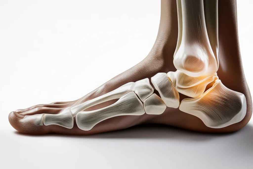
The navicular is a small, boat-shaped bone near the top of the midfoot that plays an outsized role in how the arch supports body weight. When the joints around it lose stability, navicular subluxation – a partial displacement of the bone relative to the talus, the cuneiforms, or both – can develop. It may follow an acute injury, or it may emerge gradually alongside flatfoot and posterior tibial tendon problems, making midfoot instability worse over time.
Because the navicular sits at the keystone of the medial longitudinal arch, even mild subluxation navicular bone foot instability can change how weight moves through the foot during standing, walking, and push-off. This article covers what navicular subluxation is, how it’s diagnosed, what conservative and surgical treatments the evidence supports, and where regenerative approaches may or may not fit in.
What Is Navicular Subluxation?
The navicular sits between the talus and the three cuneiform bones, forming the talonavicular joint behind it and the naviculocuneiform joint in front. Together with surrounding ligaments and the posterior tibial tendon, these joints act as a hinge for the medial arch. When soft-tissue support fails, the navicular can shift out of position – a subluxation at either the talonavicular or naviculocuneiform joint, sometimes described as naviculocuneiform “sag” (Metzl, Foot Ankle Clin, 2017).
Direct trauma, such as a fall or twisting injury, can acutely displace the navicular. More often, though, navicular bone subluxation develops gradually as part of adult-acquired flatfoot deformity, where posterior tibial tendon dysfunction allows the medial column of the foot to collapse and drift over time. Ligamentous laxity, obesity, and repetitive loading are also recognized contributors to medial column instability (Schleunes & Catanzariti, Clin Podiatr Med Surg, 2023).
Common symptoms include aching or sharp pain along the top and inner midfoot, a palpable prominence where the navicular sits, a sense that the arch is giving way, swelling over the dorsal midfoot, and reduced push-off strength because the medial column no longer transmits force efficiently. Pain often worsens with prolonged standing, uneven surfaces, or arch-loading activities like running.
Diagnosing Navicular Bone Subluxation
Diagnosis starts with a physical exam. A clinician checks for tenderness over the navicular and adjacent joints, assesses arch height sitting versus standing, and looks for the “too many toes” sign and heel-rise weakness suggesting posterior tibial tendon involvement. The navicular drop test – measuring how far the navicular tuberosity descends from non-weight-bearing to weight-bearing – is a reasonably reliable clinical tool, with a drop of roughly 10 mm or more generally considered a marker of flexible flatfoot (Zuil-Escobar et al., J Manipulative Physiol Ther, 2018).
Imaging confirms and stages the problem. Weight-bearing X-rays are the standard starting point, since they reveal alignment under normal loading – non-weight-bearing films can understate subluxation because the deformity may partially reduce once unloaded. Radiographs can show a naviculocuneiform fault, medial column elevation, or talonavicular malalignment. MRI or CT may be added to assess ligament and tendon integrity, particularly before surgical planning (Schleunes & Catanzariti, 2023).
Conventional Treatment Options
Treatment is generally staged by severity, following a framework similar to posterior tibial tendon dysfunction and adult-acquired flatfoot management.
Mild to moderate cases usually start with orthotics and activity modification. Custom or semi-custom foot orthoses that support the medial arch can help redistribute load away from an unstable navicular. Orthotic therapy is often recommended as a first-line option, even in more advanced disease stages where surgery isn’t yet indicated (Richie, Clin Podiatr Med Surg, 2020).
Bracing with an ankle-foot orthosis may be added when pain or instability is more pronounced, particularly for patients who aren’t surgical candidates, and appears to help control abnormal midfoot and hindfoot motion.
Physical therapy plays a central role at nearly every stage. Programs combining posterior tibial tendon and intrinsic foot muscle strengthening with gastrocnemius-soleus stretching have been associated with meaningful improvements in pain and function in related arch-collapse conditions (Richie, 2020). Because navicular stability depends heavily on the posterior tibial tendon and surrounding ligaments, this rehabilitation focus is evidence-supported rather than an afterthought.
Surgical stabilization is reserved for refractory or more severe navicular subluxation – persistent instability despite a genuine trial of conservative care, or progressive deformity on imaging. Procedures may include arthrodesis of the talonavicular or naviculocuneiform joint, medial cuneiform osteotomy, or tendon transfer, depending on which structures are involved (Schleunes & Catanzariti, 2023).
Regenerative options such as stem cell or platelet-rich plasma injections are sometimes discussed by patients seeking ligament and tendon support around the foot and ankle more broadly. However, dedicated clinical evidence evaluating these therapies specifically for navicular subluxation is currently limited, so they’re best viewed as a complementary consideration to raise with a specialist rather than a first-line treatment for this condition.
The Evidence: What Research Shows
Direct clinical literature on navicular subluxation as its own diagnostic entity is sparse, so much of the applicable evidence comes from the closely related fields of adult-acquired flatfoot, posterior tibial tendon dysfunction, and medial column instability – conditions in which navicular displacement is a defining feature rather than an incidental finding.
Metzl’s review of naviculocuneiform sag outlines both nonoperative and operative approaches once conservative care fails (Metzl, 2017). Schleunes and Catanzariti emphasize that medial column instability is a primary deforming force in flatfoot progression that tends to worsen rather than remain static if untreated (Schleunes & Catanzariti, 2023). Richie’s review of orthotic and biomechanical management supports arch-supporting orthoses and targeted physical therapy as reasonable first-line strategies, reserving surgery for cases that don’t respond (Richie, 2020).
Anatomic research reinforces the link between the posterior tibial tendon and the navicular: Kiter and colleagues found tibialis posterior tendon abnormalities to be common in feet with flatfoot deformity, supporting the idea that navicular instability and tendon dysfunction frequently occur together (Kiter et al., Acta Orthop Scand, 1999). Diagnostic research on the navicular drop test confirms it as a reasonably reliable measure clinicians can use to monitor arch collapse over time (Zuil-Escobar et al., 2018).
Taken together, this evidence suggests this type of midfoot displacement is best understood as part of a spectrum of instability rather than an isolated curiosity, and that staged conservative management, escalating to surgery only when needed, has reasonable support across the related literature.
Safety Considerations and Realistic Expectations
This kind of midfoot instability can progress if left unaddressed, and self-diagnosis based on foot pain alone is unreliable given how many midfoot conditions overlap in symptoms. Anyone with persistent arch pain, a visible midfoot prominence, or a sense of instability when standing or walking should be evaluated by a podiatrist or orthopedic foot and ankle specialist rather than relying on generic arch supports.
A specialist can confirm whether symptoms trace to navicular subluxation, an accessory navicular, posterior tibial tendon dysfunction, or another midfoot condition, since treatment differs meaningfully between diagnoses. Weight-bearing imaging is an important part of this evaluation. Expectations should stay realistic: conservative treatment may reduce pain and slow progression, but it doesn’t reliably reverse structural deformity once it becomes fixed, which is why earlier evaluation tends to preserve more options.
Frequently Asked Questions
What does navicular subluxation feel like?
Most people describe aching or sharp pain along the top or inner midfoot, a sense that the arch is giving way, and sometimes a visible bump where the navicular sits. Symptoms often worsen with prolonged standing or arch-loading activity.
Is navicular subluxation the same as an accessory navicular?
No. An accessory navicular is an extra bone fragment some people are born with, while navicular subluxation refers to displacement of the main bone relative to adjacent joints. The two can coexist, since an accessory navicular may weaken the posterior tibial tendon attachment and contribute to instability.
What causes subluxation of the navicular bone in the foot?
Trauma can cause it acutely, but it more commonly develops gradually alongside posterior tibial tendon dysfunction, ligamentous laxity, or long-standing flatfoot, where the midfoot progressively loses structural support.
Can this condition heal without surgery?
Many milder or earlier-stage cases are managed with orthotics, bracing, and physical therapy focused on the posterior tibial tendon and intrinsic foot muscles. Evidence suggests conservative care can reduce pain and may help slow progression, though it may not fully reverse an established deformity.
How is navicular bone subluxation diagnosed?
Diagnosis typically combines a physical exam – including the navicular drop test and posterior tibial tendon strength assessment – with weight-bearing X-rays, and sometimes MRI or CT, to confirm the degree and location of displacement.
When is surgery needed?
Surgical stabilization, such as joint fusion or osteotomy, is generally considered when pain and instability persist despite an adequate trial of conservative treatment, or when imaging shows progressive deformity.
Can stem cell therapy treat navicular subluxation?
Evidence evaluating regenerative therapies specifically for this condition is currently limited. These options are sometimes explored as a complementary consideration for ligament and tendon health around the foot and ankle, but they aren’t established as primary treatment for this condition and should be discussed with a specialist alongside conventional care.
Key Takeaways
- Navicular subluxation involves partial displacement of the navicular bone at the talonavicular or naviculocuneiform joint, disrupting the medial arch’s structural support.
- Causes range from acute trauma to gradual instability associated with posterior tibial tendon dysfunction, ligamentous laxity, and long-standing flatfoot.
- Common symptoms include midfoot pain, a visible prominence, arch collapse, and reduced push-off strength.
- Diagnosis relies on exam findings such as the navicular drop test, plus weight-bearing X-rays and sometimes MRI or CT for surgical planning.
- Orthotics, bracing, and physical therapy targeting the posterior tibial tendon and intrinsic foot muscles are generally the first-line approach for milder cases.
- Surgical stabilization is reserved for refractory or progressive cases that don’t respond to conservative care.
- Current evidence for regenerative therapies in this specific condition is limited, so they should be viewed as complementary rather than primary treatment.
- Early evaluation by a podiatrist or orthopedic specialist helps preserve treatment options before deformity becomes structurally fixed.
Anyone with persistent midfoot pain or arch instability should consult a qualified podiatrist or orthopedic specialist for a proper diagnosis; those exploring regenerative options for ligament and tendon support as part of a broader care plan can schedule a consultation with Ways2Well or learn more about regenerative medicine beyond stem cells.
For related reading, see Ways2Well’s guides on stem cell injections for foot pain and stem cell therapy for ankle injuries.
References
- Metzl JA. “Naviculocuneiform Sag in the Acquired Flatfoot: What to Do.” Foot and Ankle Clinics, 2017;22(3):529–544. DOI: https://doi.org/10.1016/j.fcl.2017.04.007
- Schleunes S, Catanzariti A. “Addressing Medial Column Instability in Flatfoot Deformity.” Clinics in Podiatric Medicine and Surgery, 2023;40(2):271–291. DOI: https://doi.org/10.1016/j.cpm.2022.11.003
- Richie DH. “Biomechanics and Orthotic Treatment of the Adult Acquired Flatfoot.” Clinics in Podiatric Medicine and Surgery, 2020;37(1):71–89. DOI: https://doi.org/10.1016/j.cpm.2019.08.007
- Zuil-Escobar JC, Martínez-Cepa CB, Martín-Urrialde JA, Gómez-Conesa A. “Medial Longitudinal Arch: Accuracy, Reliability, and Correlation Between Navicular Drop Test and Footprint Parameters.” Journal of Manipulative and Physiological Therapeutics, 2018;41(8):672–679. DOI: https://doi.org/10.1016/j.jmpt.2018.04.001
- Kiter E, Erdag N, Karatosun V, Günal I. “Tibialis posterior tendon abnormalities in feet with accessory navicular bone and flatfoot.” Acta Orthopaedica Scandinavica, 1999;70(6):618–621. DOI: https://doi.org/10.3109/17453679908997852
Author: Ways2Well Editorial Team
Reviewed by: Scientific Advisory Board member